Dr. Cristina Pérez, dermatologist at the HLA Toledo Medical Center
“Digital dermatoscopy aids the early diagnosis of melanoma”
Thanks to digital dermatoscopy the shape, size, colour or symmetry of marks on the skin can be analysed. But, beyond the fact that it is a non-invasive examination, what benefits does it bring for both patients and doctors?
Patients with digital dermatoscopy monitoring have melanomas detected in earlier stages and it helps to detect changes that are more subtle than when using a normal examination. As a result of this, we can remove suspicious lesions much earlier, minimising unnecessary surgery. Additionally, it allows a very detailed monitoring of the moles, particularly in patients with a personal or family background of atypical pigmented lesions and melanomas.
What does this technique really involve?
The idea is to design a body map of lesions by zones using normal photography of each patient and a dermatoscopic image of the lesions to be monitored with their evolution over time. Digital dermatoscopy allows the lesions to be photographed with great diagnostic accuracy, filing them and comparing the images of the same lesion over time, aiding its monitoring. The dermatologist takes general images of the patient and then dermatoscopic images of all the marks to be monitored, around 30-40 lesions. In the following check-ups, the evolution of these images is stored and compared.
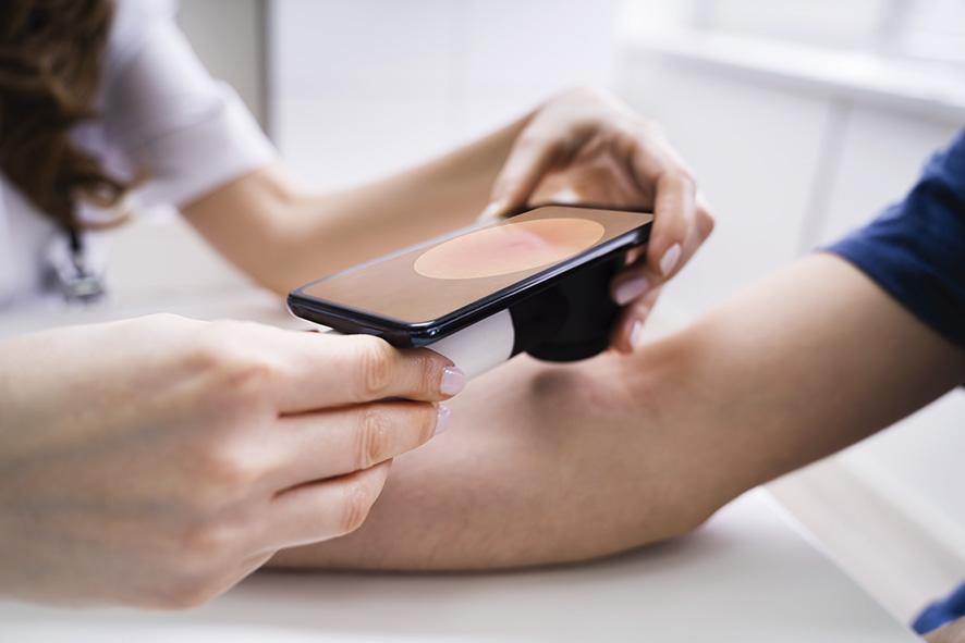
And how are these photographs taken?
Digital dermatoscopy is a non-invasive technique that allows the analysis of structures that are not visible in a normal physical examination. To do this, a magnifying glass and a lighting system that makes the cornea layer of the epidermis translucent are used. The dermatoscopy analyses and stores a detailed image of the moles, generating a map of our skin.
Could you explain a case where it is highly beneficial?
Melanoma is the most aggressive skin cancer that exists and its removal in initial phases is a determining factor in the good prognosis of patients diagnosed with this tumour. Regular monitoring using digital dermatoscopy of atypical lesions allows, in most cases, an early detection of the melanoma. Digital dermatoscopy increases the probabilities of early diagnosis of a melanoma by 60-90%.
This highly detailed observation is useful for existing marks, but can it help prevention?
Yes, naturally. It does not only allow us to see the evolution of the dermatoscopic lesion, but rather it gives us a map of the patient’s body using normal photography, where it is possible to detect new lesions.
Who she is At the age of 41 years, Doctor Cristina Pérez became the youngest head of a Dermatology service in Toledo in the Spanish public health system. Married, with three daughters, for this doctor the only secret of a job well done is: effort and sacrifice.
She studied Medicine at the Universidad Autónoma de Madrid; she prepared the MIR (medical residency examination) in Oviedo and, with ranking number 558, she chose her Dermatology residency in Toledo. When she was 31 years old, she gained her position as medical specialist in Dermatology, and ten years later, she became head of her area.